Rebleeding
The onset of rebleeding is usually accompanied by sudden severe headache, often associated with severe nausea and vomiting. Rebleeding can be confirmed by a CT scan or a sudden spike in ICP with new blood seen in the bag if a ventricular drain is in place. Early treatment, with either surgical or endovascular methods, of the aneurysm is the most effective means of preventing rebleeding.
Cerebral Vasospasm
By definition, cerebral vasospasm is narrowing of a cerebral blood vessel and causes reduced blood flow distally, which may lead to delayed ischemic deficit and cerebral infarction if left untreated. Besides the damage done by the initial SAH, brain damage produced by vasospasm is an important cause of morbidity and mortality after hemorrhage.
Hydrocephalus
Hydrocephalus is a condition in which there is either an obstruction to the flow of CSF within the ventricular system or subarachnoid space either due to intraventricular mass lesions or to external compression or a problem with reabsorption of CSF. Hydrocephalus can be classified as acute, subacute, or delayed. With SAH, hydrocephalus develops as a result of blood in the CSF, thus interfering with the reabsorption of CSF.
Seizures
Risk factors for seizures in the early period after SAH include previous history of hypertension, CT-documented presence of focal intraparenchymal blood, occurrence of a cerebral infarction, middle cerebral aneurysm location, and duration of coma after SAH. If the hemorrhage is mild, anticonvulsants are tapered after 1 month. If the hemorrhage is more severe, extended therapy and EEG monitoring are employed.

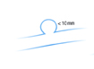 Small aneurysms are less than 10 mm in diameter
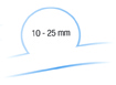 Large aneurysms are 10-25 mm in diameter
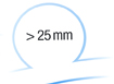 Giant aneurysms are greater than 25 mm in diameter
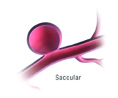 Saccular cerebral aneurysms are intracranial aneurysms with a characteristic rounded shape
 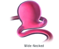 Wide-Necked aneurysms are Saccular cerebral aneurysms with a characteristic wide-necked shape
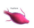 The fusiform aneurysm looks like an outpouching of an arterial wall on both sides of the artery.